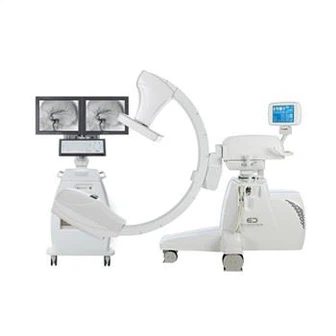
The Medical Angiography X-ray System
The Medical Angiography X-ray System enhances the signal of the uncontrasted image after passing through the human body, and then uses a high-resolution camera to scan the enhanced image. The digital information of the contrast image and the digital information of the uncontrast image are subtracted, and the difference between the two is expressed as different gray levels, and the difference image is formed on the display. Since the bone and soft tissue images of the two images have the same value, they are eliminated in the subtraction, leaving only the blood vessel image containing the contrast agent. As a result, structures other than the contrast-enhanced blood vessel are eliminated, and the image of the organ being contrasted is highlighted. The image signal after subtraction processing is proportional to the thickness/density of the contrast agent, is related to the absorption coefficient of the contrast agent and blood vessels, and has nothing to do with the background.