Understanding the Subtraction Process in Digital Subtraction Angiography
Digital Subtraction Angiography (DSA) is a critical imaging technique in medical diagnostics, particularly for visualizing blood vessels. One of its key innovations is the subtraction process, which significantly enhances image clarity by isolating vascular structures from surrounding tissues and bones. This article explains the subtraction process in simple terms to make it accessible to medical professionals and curious readers alike.
What is DSA?
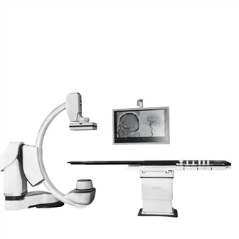
DSA is a specialized X-ray technique used to visualize blood vessels after injecting a contrast agent. Unlike traditional angiography, DSA employs digital technology to create highly detailed and focused images of the vascular system. This makes it invaluable for diagnosing vascular diseases, planning surgeries, and guiding interventional procedures.
How the Subtraction Process Works
The subtraction process in DSA involves three main steps:
Acquiring the Mask Image: A mask image is taken before the injection of the contrast agent. This image captures the background structures, including bones, soft tissues, and other anatomical features that could obscure the blood vessels.
Acquiring the Contrast Image: After the contrast agent is injected, a second X-ray image is taken. The contrast agent fills the blood vessels, making them more visible on the image.
Image Subtraction: The mask image is digitally subtracted from the contrast image. This process removes the background structures, leaving only the enhanced blood vessels in the final image. The resulting clarity allows for precise analysis of the vascular system.
Advantages of the Subtraction Process
Enhanced Visualization: By removing background structures, the subtraction process highlights even small or intricate blood vessels.
Reduced Need for Multiple Angles: Clearer images often eliminate the need for acquiring images from multiple angles.
Minimized Contrast Agent Use: Precise imaging reduces the volume of contrast agent required, which is beneficial for patients with kidney issues.
Applications in Medicine
The subtraction process is used in various medical fields, including:
Neurology: For diagnosing aneurysms or vascular malformations.
Cardiology: To assess coronary artery diseases.
Interventional Radiology: To guide procedures such as angioplasty or stent placement.
Challenges and Considerations
While the subtraction process is highly effective, it requires:
Patient Stability: Any movement between the mask and contrast image acquisition can lead to artifacts and reduce image quality.
High-Quality Equipment: Advanced imaging systems and software are essential for accurate subtraction.
Experienced Operators: Technicians and radiologists must be well-trained to optimize the process and interpret the images.
Conclusion
The subtraction process in DSA revolutionized vascular imaging, providing unparalleled clarity and detail. By isolating blood vessels from other anatomical structures, this technique has become an indispensable tool in modern medicine, aiding in accurate diagnoses and effective treatments. As technology continues to evolve, further enhancements in the subtraction process promise even greater benefits for patients and healthcare providers alike.