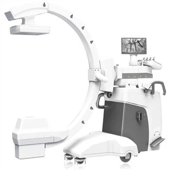
Specific Operation Of Angiography Technology
In order for the inspection to be completed under the best sanitary conditions, you must be at the side of the examination table and place a disinfection sheet on the examination table. During the entire inspection process, nurses and doctors must be present. After local anesthesia in the groin, a fine needle is inserted into the artery. The guide wire is inserted into the blood vessel through a fine needle. The role of the guide wire is to guide the synthetic catheter to the desired position under exposure. By using a catheter to inject iodine-containing contrast media, the blood vessels of different organs can be displayed. After removing the catheter, use a bandage and dressing to compress the puncture site to stop bleeding.
Angiography usually refers to digital substraction angiography (digital substraction angiography, DSA) is a technology that uses computers to process digital image information to eliminate bone and soft tissue images and make blood vessels clear. Nuldelman obtained the first DSA image in 1977, which has been widely used in clinics, replacing the older generation of non-subtraction angiography methods. In recent years, the development of tablet DSA has become faster and faster, and the image quality and the safety of doctors and patients have also been continuously improved. A new generation of diagnostic equipment represented by the Siemens Artis zee series digital tablet DSA has emerged to provide clinical cardiovascular diagnosis and treatment. New digital technology.
The imaging methods of DSA are divided into intravenous injection digital subtraction angiography (IVDSA) and arterial injection digital subtraction angiography (IADSA). The former refers to the placement of a catheter or trocar injection of contrast agent through the intravenous route for DSA examination, which can be divided into non-selective IVDSA, that is, catheter placement in the peripheral vein or superior vena cava to display arterial images, and selective IVDSA, that is, catheter head placement Contrast agent is injected into the vein or heart cavity for imaging. The latter can also be divided into non-selective angiography and selective angiography. Non-selective IADSA means that the tip of the catheter is placed in the proximal aorta of the target artery to inject contrast agent for antegrade visualization after puncturing the catheter through the arterial route; while selective IADSA means that the tip of the catheter is further deep into the target artery Perform radiography within the main trunk or branches of the main trunk.