Medicine and Equipment for Making Cardiovascular angiography
Selective cardiovascular angiography requires a suitable contrast agent and X-ray rapid continuous photography equipment, as well as a suitable cardiac catheter and pressurized syringe for angiography.
The contrast agent is 60-76% diatrizoate meglumine, and the dose is: 1 to 1.5ml/kg per time for children, and the total dose shall not exceed 4ml/kg; 50-60ml per time for adults, and the total dose does not exceed 200ml. An iodine allergy test is required before use. 1ml of 30% diatrizoate meglumine can be used as an intravenous injection. Observe for 20 minutes and use it after no response. If you are allergic to diatrizoate meglumine, you can use a non-ionic contrast agent, such as triiodotriphenyl, it may also produce some mild reactions such as nausea, vomiting, skin itching and rash in a small number of patients, so there is a history of iodine allergy Patients should also consider emergency treatment in advance, or use corticosteroids and antihistamines before imaging. In addition to the low risk of response, non-ionic contrast agents produce better image effects, but they are expensive and have not been widely used.
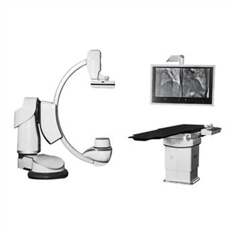
The X-ray machine used for cardiovascular imaging needs to output a large number of X-rays in a short time, thereby shortening the exposure time, facilitating rapid continuous photography and improving image clarity, so a large capacity above 50mA is required. The power of X-ray tube is above 50kW. For simultaneous shooting in both directions, an X-ray machine above 1000kW or two X-ray machines above 500kW are required for simultaneous shooting.
Fast photography requires a fast film changer to change the film during continuous exposure. Generally, the rapid film changer can take up to 6 films per second, and the two film changers in the front and side positions can be synchronized. Fluorescence image enhancement device and X-ray film photography can be used to take 24-80 frames of images per second. The entire imaging process can be monitored by the TV system. The flow of contrast agent in the heart, blood vessels, and various parts of the heart and great blood vessels can be monitored during film screening. The structure is dynamically observed. At the same time, the radiography process can also be recorded on tape.
Choose the front, side, or left and right oblique positions when shooting as needed, and in recent years, use the axial angle projection. Make the solution part of the radiography show better results.
Digital subtraction angiography is a comprehensive image enhancement-TV system data collection and computer processing to generate images, which can significantly reduce the concentration and dose of contrast agent, and receive excellent diagnostic results, thereby reducing or avoiding high-concentration, large-scale contrast agents The side effects during injection are especially suitable for cases with reduced renal function.
Cardiac catheters for imaging require a large lumen so that the contrast agent can pass quickly. Adults commonly use F8 and F9 imaging catheters, and children can use F6 and F7 catheters. When the contrast agent is injected into the large blood vessel cavity of the heart through the cardiac catheter, it is required to form a pellet, and enter the blood quickly from the front end of the cardiac catheter to achieve the highest concentration and clear visualization in this part. Therefore, the injection must be fast. Because the contrast agent has a certain viscosity and the catheter cavity has a certain resistance, it is necessary to use the Kamura syringe to achieve the purpose of rapid injection. Generally, a dose of medicine is injected in about 1.5 seconds. After a certain amount of contrast agent is injected, the syringe is triggered The exposure trigger device for rapid filming or cinematography to start photography. If it is required to inject at a certain time in the cardiac cycle, a certain waveform of the patient's electrocardiogram must be used as a signal to trigger the injector to start.