Coronary Angiography: A Clear Look at Your Heart’s Arteries
Coronary angiography is a highly effective and commonly used procedure for diagnosing coronary heart disease (CHD), a condition in which the heart's own blood vessels become narrowed or blocked.
Often called "cardiac catheterization," this minimally invasive test allows doctors to directly visualize the inside of your coronary arteries.
How Does It Work?
Accessing the Artery: The procedure begins with a doctor making a small puncture, usually in an artery in the groin or wrist. A thin, flexible tube called a catheter is gently inserted.
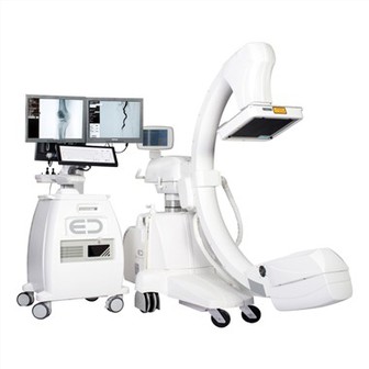
Guiding to the Heart: Using live X-ray imaging as a guide, the doctor carefully navigates the catheter through the body's main blood vessel (the aorta) until it reaches the coronary arteries.
Taking the Images: A special dye (contrast agent) is injected through the catheter directly into the coronary arteries. As the dye flows through the vessels, X-ray videos are recorded. The dye outlines the arteries clearly, revealing any narrowings or blockages caused by plaque buildup.
Why Is It Important?
Images from coronary angiography provide a detailed "roadmap" of the heart's blood supply. This allows doctors to:
Identify the precise location of any blockages
Assess the extent and severity of the narrowing
Determine the best treatment plan-whether placing a stent, recommending bypass surgery, or managing the condition with medication