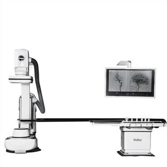
Cardiovascular Angiography
Cardiovascular angiography is a medical imaging technique that involves the rapid injection of a transparent contrast agent containing organic compounds into the bloodstream under X-ray irradiation. This procedure is designed to visualize the heart and large blood vessel cavities, providing clear and detailed imaging of cardiovascular structures.
During the process, auxiliary methods such as rapid imaging, fluoroscopy, or video recording are utilized to capture the dynamic visualization of the heart and major blood vessels. These techniques allow medical professionals to record the blood flow sequence containing the contrast agent and observe the filling patterns of the heart's large vessels in real-time. This dynamic process is crucial for identifying abnormalities in blood flow or vascular structure.
From the imaging results, physicians can assess both physiological and anatomical changes in the cardiovascular system. For example, they can detect blockages, aneurysms, malformations, or changes in cardiac function. Additionally, angiography provides critical information for planning therapeutic interventions, such as angioplasty or bypass surgery, and for monitoring the success of previous treatments.
As one of the most reliable diagnostic tools in cardiovascular medicine, angiography offers unparalleled insights into the functional and structural aspects of the heart and blood vessels. Its high diagnostic value makes it an essential procedure in identifying, managing, and treating cardiovascular diseases.